LONG CASE: FINAL PRACTICAL (1701006166)
MEDICAL CASE
This is an E log book to discuss our patient's de-identified health data shared after taking his guardian's signed informed consent. Here we discuss our individual patient problems through series of inputs from available global online community of experts with an aim to solve those patient's clinical problems with collective current best evidence based inputs. This e-log book also reflects my patient centered online learning portfolio and your valuable comments in comment box are most welcomed
I have been given this case to solve in an attempt to understand the topic of "Patient clinical data analysis" to develop my competency reading and comprehending clinical data including history, clinical findings, investigations and come up with a diagnosis and treatment plan.
SARBESH MISHRA
Hall ticket No:1701006166
CASE DISCUSSION:
- Shortness of breath since 7 days
- Chest Pain on left side since 5 days
- Insidious in onset
- Gradually progressive (From grade I to grade II according to mMRC scale)
- Aggravates on exertion and Postural variation (i.e lying on left lateral side it increases)
- Relieved on rest and sitting position.
- Chest pain: non radiating, nature is pricking type
- loss of weight (approx 10kgs in past 1yr)
- loss of appetite
- Vomitings
- Orthopnea, PND (paroxysmal nocturnal dyspnea)
- Edema
- Palpitations
- Wheeze
- Chest tightness
- Cough
- Hemoptysis
- HTN
- ASTHMA
- CAD
- EPILEPSY
- TB.
- Diet- Mixed
- Sleep- Adequate
- Appetite- Decreased
- Bowel and bladder- Regular
- Alcohol- Stopped 20years back (Before 90ml per day)
- Smoking- From past 20years (10 cigarettes per day), stopped 2years back
- No known allergies
Patient is conscious, coherent and co-operative.
He is well oriented to time, place and person.
Moderately built and nourished.
No signs of pallor, edema, icterus, cyanosis, clubbing, lymphadenopathy.
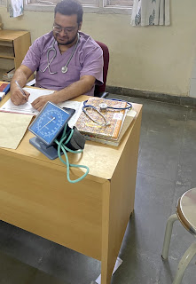
VITALS:
- Temperature : Afebrile
- Pulse rate : 139beats/min
- BP : 110/70 mm Hg
- RR : 45 cpm
- SpO2 : 91% at room air
- GRBS : 201mg/dl
.jpeg)


.jpeg)

.jpeg)
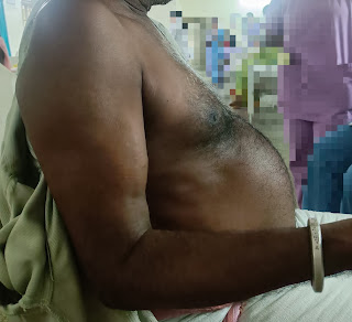
INSPECTION:
Shape of chest is elliptical,
B/L asymmetrical chest,
Expansion of chest- Right- normal, left-decreased.
PALPATION:
All inspectory findings are confirmed,
No tenderness, No local rise of temperature,
trachea is deviated to the right,
Measurement:
AP: 24cm
Transverse:28cm
Right hemithorax:42cm
left hemithorax:40cm
Circumferential:82cm
Tactile vocal fremitus: decreased on left side ISA, InfraSA, AA, IAA.
PERCUSSION:
AUSCULTATION:
B/L air entry present, vesicular breath sounds heard,
Decreased intensity of breath sounds in left SSA,IAA,
Absent breath sounds in left ISA.
2. CVS EXAMINATION:
S1,S2 heard
No murmurs. No palpable heart sounds.
JVP: normal
Apex beat: normal
Soft, Non-tender
No organomegaly
Bowel sounds heard
No guarding/rigidity
4. CNS EXAMINATION:
No focal neurological deficits
Gait- NORMAL
Reflexes: normal
DB: 0.74mg/dl
AST: 24IU/L
ALT: 09IU/L
ALP: 167IU/L
TP: 7.5gm/dl
ALB: 3.29gm/dl

.jpeg)

.jpeg)
- Moderate Pleural effusion in left lobe of lungs.
- Right sided lung consolidation.


- O2 inhalation with nasal prongs with 2-4 lt/min to maintain SPO2 >94%
- Inj. Augmentin 1.2gm/iv/TID
- Inj. Pan 40mg/iv/OD
- Tab. Pcm 650mg/iv/OD
- Syp. Ascoril-2tsp/TID
- DM medication taken regularly
- High Protein diet
- 2 egg whites/day
- Monitor vitals
- GRBS every 6th hourly
SARBESH MISHRA
Hall ticket No:1701006166
Comments
Post a Comment